




 Download PDF
ABSTRACT
Background
Preeclampsia is a pregnancy-specific hypertensive disorder characterized by the onset of hypertension and proteinuria after the 20th week of gestation in previously normotensive women. It remains a leading cause of maternal and fetal morbidity and mortality worldwide. Delivery of the placenta is currently the only definitive treatment, underscoring its central role in disease pathogenesis.
Objective
This review aims to summarize and integrate current knowledge on the molecular mechanisms underlying the development of preeclampsia, with a focus on placental dysfunction and maternal vascular responses.
Methods
A narrative review of the literature was conducted, focusing on studies investigating the molecular and cellular basis of preeclampsia, including angiogenic imbalance, oxidative stress, inflammation, and dysregulation of vascular signaling pathways.
Results
Placental ischemia, resulting from inadequate trophoblast invasion and insufficient remodeling of maternal spiral arteries, is recognized as a key initiating factor. This leads to reduced uteroplacental perfusion and hypoxia. In response, the ischemic placenta releases a range of bioactive factors into the maternal circulation that promote systemic endothelial dysfunction. These factors contribute to oxidative stress, activation of inflammatory pathways, dysregulation of the renin–angiotensin system, and an imbalance between pro-angiogenic and anti-angiogenic mediators. Collectively, these alterations impair endothelial function, disrupt vascular homeostasis, and result in hypertension and end-organ damage.
Conclusion
Preeclampsia arises from complex and interconnected molecular mechanisms involving placental dysfunction and maternal endothelial injury. A deeper understanding of these pathways may facilitate the identification of novel biomarkers and therapeutic targets, ultimately improving the prevention and management of this disorder.
Keywords: Preeclampsia; Placental ischemia; Endothelial dysfunction; Oxidative stress; Angiogenic imbalance; Inflammation; Renin–angiotensin system
INTRODUCTION
Preeclampsia remains a major contributor to maternal morbidity, mortality, and preterm birth worldwide [1]. It is characterized by the new onset of hypertension (≥140/90 mmHg) and proteinuria (≥0.3 g in a 24-hour urine collection) after 20 weeks of gestation in a previously normotensive woman. The condition most commonly affects healthy nulliparous women. However, multiparous women with a new partner are also at increased risk, suggesting an immunological component in its pathogenesis. Furthermore, a history of preeclampsia in a prior pregnancy significantly increases the likelihood of recurrence in subsequent pregnancies. Severe forms of the disease may progress to eclampsia, defined by the occurrence of seizures, and/or HELLP syndrome, a life-threatening complication characterized by hemolysis, elevated liver enzymes, and low platelet count, reflecting multisystem involvement [2].
Termination of the pregnancy is the only effective treatment for preeclampsia, as the onset and progression of the disorder are unpredictable. Preeclampsia may also occur in women with abdominal pregnancies. It is not likely, or at least not necessary, that the uterus plays a part. Evidence shows that preeclampsia is a two-stage disorder. It derives primarily from a placental ischemia, secondarily through alteration of endothelial function [3]. During pregnancy, the trophoblast invades and remodels the uterine spiral to increase the blood flow into the intervillous space and invasion of the placenta. Cytotrophoblast invasion of the uterine wall leads to an unusual ectoderm to vascular mesoderm transformation by adhesion molecule switching. Cytotrophoblast progenitors, which express adhesion molecules, are characteristic of epithelial cells. In preeclampsia, however, this process failed to develop. Inadequate cytotrophoblast invasion in preeclampsia causes reduced utero-placental perfusion, which in turn leads to hypoxia. The ischemic placenta releases toxic factors into the maternal circulation, which leads to an exaggeration of the systemic maternal inflammatory response [4]. Molecular mechanisms contribute to the disease process. Among these altered angiogenic balance, systemic inflammation, oxidative stress, alteration of the renin-angiotensin system, and placental hypoxia and ischemia play a part; it is unknown whether the mechanisms act together or independently [5].
METHODOLOGY
This study was conducted as a narrative review aimed at synthesizing current knowledge on the molecular basis of preeclampsia. A flexible and interpretive approach was adopted to integrate findings from diverse areas of research, including molecular biology, vascular physiology, and placental pathology.
A comprehensive literature search was performed using electronic databases, including PubMed/MEDLINE, Scopus, Web of Science, and Google Scholar. The search focused on articles published between January 2000 and December 2025. Keywords and Medical Subject Headings (MeSH) terms were used in various combinations, including “preeclampsia,” “molecular mechanisms,” “pathophysiology,” “angiogenic factors,” “VEGF,” “PlGF,” “endothelial dysfunction,” “oxidative stress,” “inflammation,” and “renin–angiotensin system.” Relevant references cited within selected articles were also reviewed to identify additional sources.
Articles were selected based on their relevance to the molecular and pathophysiological mechanisms of preeclampsia. Priority was given to peer-reviewed original research articles, review papers, and meta-analyses that provided significant insights into disease mechanisms. No formal systematic inclusion or exclusion criteria were applied; however, studies focusing solely on clinical management without mechanistic relevance were generally excluded.
The selected literature was critically reviewed and thematically organized into key areas, including abnormal placentation, angiogenic imbalance, oxidative stress, inflammatory pathways, and dysregulation of the renin–angiotensin system. Findings were synthesized descriptively to provide a coherent overview of the current understanding of the molecular mechanisms involved in preeclampsia.
Ethical Considerations
As this study is a review of previously published literature, ethical approval was not required.
Placental Hypoxia
The circulatory demands increase during pregnancy. It places significant stress on the maternal cardiovascular system. It also increases the blood volume by half. Cardiac output and blood flow to the uterus increase by 40% and eightfold, respectively. This causes the maternal vascular system to reform itself for the fetus and placenta to receive adequate oxygen and nutrients, and for the pregnancy to proceed [6].
The maternal spiral arteries remodel themselves from high-resistance vessels rich in smooth muscle to dilated, high-capacitance vessels by the invasion of extravillous trophoblasts of fetal origin. However, a pre-eclamptic placenta does not develop normally. Trophoblasts do not differentiate properly and are unable to invade the myometrium effectively. Spiral artery remodeling is limited and persists as higher resistance vessels, which results in less blood flow. This results in placental ischemia, though it is unclear whether it is a cause (ischemia causing the placenta to release factors) or a result (arteries that fail to dilate, causing ischemia) of abnormal placentation. The muscular, narrow spiral vessels are unable to meet the increased demands. Placental ischemia will occur throughout the pregnancy period [7]. The hypoperfusion leads to the release of many factors into the maternal circulation, such as antiangiogenic factors, inflammatory mediators, immune cells, hypoxia-inducing factor 1a, and angiotensin-1 autoantibodies. As a result of the release of these factors, it causes excessive inflammatory response and an increased oxidative environment within the vasculature. It induces endothelial activation and dysfunction, which is characterized by increased vasoconstriction, decreased vasodilatation, and altered angiogenesis. This imbalance increases maternal blood pressure and peripheral vascular resistance and results in a negative feedback loop, which will exacerbate ischemic insult to the placental environment [8].
As a result of this ischemic attack, HIF-1a will be released, which is a subunit of the heterodimeric transcription factor, hypoxia inducible factor 1 (HIF-1). It is responsible for regulating the cellular response to low oxygen tension. Its expression is increased in preeclampsia and upregulates angiogenic proteins such as soluble fms-like tyrosine kinase 1 (sFlt-1), soluble Endoglin (sEng), and Endothelin-1, which gives an insight that hypoxia inducible factor 1a (HIF-1a) is a possible mediator between the placenta and vasoactive molecules that damage the maternal vascular wall. It also expresses other angiogenic factors, including vascular endothelial growth factor (VEGF), placental growth factor (PlGF), and VEGFR 1 and transforming growth factor β3 (TGF-β3), which block cytotrophoblast invasion [9].
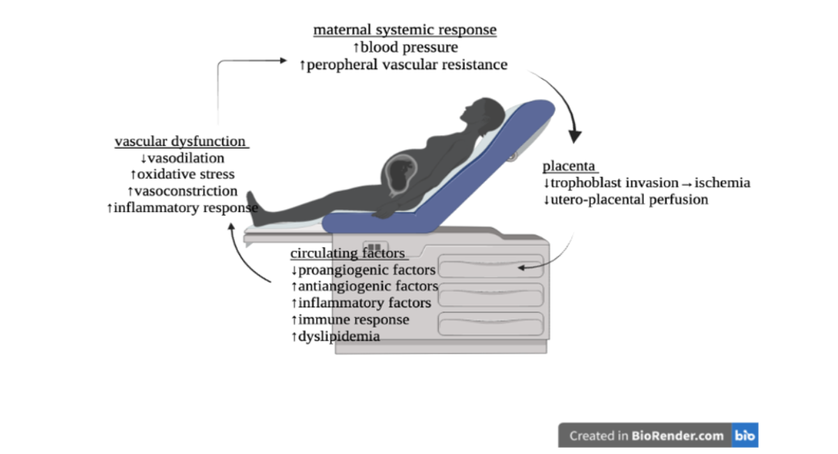
Figure 1: Factors involved in the progression of preeclampsia
The hypoxic condition also affects the down-regulator of hypoxia inducible factor 1-a, such as Catechol-O-methyltransferase (COMT). COMT metabolizes estradiols to 2-methyl estradiol (2-ME). 2-ME maintains oxygen balance and suppresses placental hypoxia, HIF-1α expression, and sFlt1 expression. The exact mechanism by which 2-ME prevents PE (preeclampsia) remains unclear. Recently, research shows that 2-ME participates in cytotrophoblast invasion. This indicates that 2ME could prevent the pathogenesis of PE by enhancing normal placental vascular formation [10].
Alteration of Angiogenic Balance in Preeclampsia
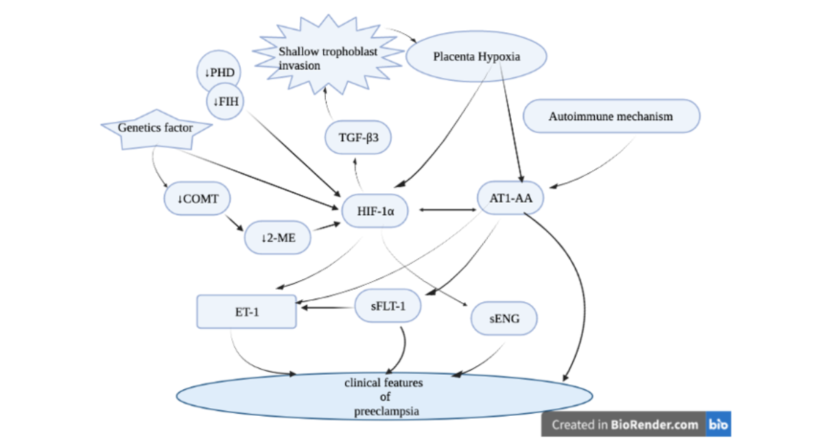
Figure 2: Role of hypoxia inducible factor 1a in the pathogenesis of preeclampsia
Alteration of Angiogenic Balance in Preeclampsia
During normal pregnancy, placental development, cytotrophoblasts (CTBs) invade the myometrium. Invading CTBs upregulate the expression of molecules that are central to uterine invasion and pseudo-vasculogenesis (the process by which CTBs switch their adhesion molecules to mimic those of vascular cells). It enhances angiogenesis and promotes the expression of angiogenic molecules from the VEGF family. Invasive CTBs express VEGF-A, VEGF-C, PlGF, VEGFR-1, and VEGFR-3. The interactions among these molecules are critical for invasion and pseudo-vasculogenesis. This leads the maternal uterine spiral arteries to remodel and transform themselves into low-resistance, high-capacitance vessels. However, this process does not happen in preeclampsia, and the expression of angiogenic factors is down-regulated [11]. As a consequence of placental hypoxia, a variety of angiogenic factors are produced by the human placenta, particularly vascular endothelial growth factor (VEGF) and placental growth factor (PlGF) (12).
VEGF is an endothelial-specific mitogen that stimulates proliferation, survival, migration, and new vessel formation, and stabilizes the endothelium in mature blood vessels. It is also a potent vascular permeabilizing agent and a vasodilator (13). It has two high-affinity receptor tyrosine kinases, VEGF receptor-1 [also called fetal liver tyrosine-like (Flt-1)] and VEGF receptor-2 [also called kinase domain-related receptor (KDR). When VEGF receptor-2 is activated, it induces endothelial cells to release NO and PGI2, which promote endothelium-dependent relaxation [14]. PlGF, a potent angiogenic growth factor, has structural homology to VEGF-A. It enhances the VEGF signaling by displacing VEGF from the Flt1 receptor and allowing it to bind to the more active kinase insert domain (KDR) receptor (or VEGFR-2). Under conditions of ischemia, inflammation, and wound healing, PlGF induces angiogenesis. Inhibition of both PlGF and VEGF could alter angiogenesis and participate in the pathogenesis of preeclampsia [15].
VEGF receptors are localized to caveolae and participate in cell signaling and transport. It initiates a signaling cascade upon phosphorylation and results in angiogenesis [16]. The hypoxic placenta releases trophoblast fragments to maternal circulation, including the antiangiogenic factors, soluble fms-like tyrosine kinase 1 (sFlt-1) and soluble Endoglin (sEng). These factors trap circulating vascular endothelial growth factor (VEGF), placental growth factor (PlGF), and transforming growth factor β (TGFβ), and prevent interaction with their endogenous receptors, decreasing their free levels respectively. This results in endothelial dysfunction and the appearance of clinical manifestations [17].
FLT-1(fetal liver tyrosine-like kinase) is a membrane-spanning receptor for VEGF and PlGF. Through differential mRNA processing, FLT-1(fetal liver tyrosine-like kinase) generates Fms-like tyrosine kinase 1 (Flt-1), and soluble Flt-1 (sFlt-1). It has an extracellular domain, a membrane domain, and a cytoplasmic tyrosine kinase domain. Soluble Flt-1 is a shorter isoform that has only the extracellular ligand-binding domain, which is secreted into the circulation. Another mechanism to generate soluble Flt-1 is through cleavage of the membrane receptor by proteases, but the physiologic role of this process is not known. Soluble Flt-1 binds VEGF and PlGF in circulation and results in preventing them from interacting with their membrane receptors on the endothelium. This leads to decreased free and bioactive PlGF and VEGF [18,19].
sFlt-1 contains heparin-binding domains that account for its strong avidity to the heparin sulfate on cell surfaces or on the extracellular matrix. Heparanase releases sFlt-1 that is retained in ligand-binding extracellular storage. In contrast, placental alteration in the expression of heparanases is mild. This chemical property raised questions about the mechanism by which Flt-1 gains access to the maternal circulation [18,20]. Release of trophoblast-derived material/fragments into the maternal circulation is the main source of sFlt-1 in preeclampsia pathogenesis. Besides, physiological function is an unclear phenomenon [21]. Other factors released by the placenta as a result of the physiologic alteration are endoglin. It is one of the co-receptors for transforming growth factor- 1 and - 3. Vascular endothelial cells and syncytiotrophoblasts highly expressed it. Release of sEng to the maternal circulation results in interference with TGFb signaling and eNOS activation, which causes endothelial dysfunction. Placental endoglin level increased in preeclampsia [22].
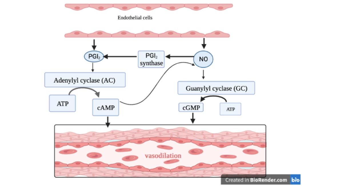
Figure 3: PGI and NO mediated vasodilatation
The Role of Nitric Oxide in Preeclampsia
NO mediates vasodilation of the endothelium, and it is responsible for the regulation of the decrease in peripheral vascular resistance in pregnancy. VEGF induces nitric oxide and prostacyclins. They promote vasodilation. As VEGF is down-regulated in preeclampsia, the availability of bioactive NO will decrease. This implies decrease NO may contribute to the pathogenesis of hypertension in preeclampsia. Additionally, oxygen-free radicals might alter endothelial cell function by scavenging NO. It halts the bioavailability of this potent vasodilator. Besides, reduced NO may result in an increase in plasma concentration of S-nitrosoalbumin in preeclampsia, which is secreted in a low level of NO state. Since S-nitrosoalbumin is responsible for promoting relaxation of resistance-sized vessels is enhanced by ascorbate. This decreased release of NO may be due to decreased vitamin C levels [23,24].
Another mechanism that might contribute to reducing NO levels involves arginase, which is involved in the conversion of L-arginine to L-ornithine and urea. NO synthase (NOS) catalyzes the conversion of L-arginine to L-citrulline and NO [25]. Since L-arginine is a common substrate for NOS and arginase, arginase reciprocally regulates NOS bioavailability. As the activity of arginase increases, it results in decreased NO production. Moreover, a reduction in the substrate concentration could result in uncoupling of endothelial NOS (eNOS), whereby electron flow from the reductase domain to the oxygenase domain is diverted to molecular oxygen rather than to L-arginine, resulting in the generation of superoxide anion. This shows a decrease in the generation of NO as well as increased scavenging of NO by superoxide to generate peroxynitrite [26].
Arginase has two isoforms (arginase I and II) that are localized in the cytosol and mitochondria, respectively. Preeclampsia shows an increase in mRNA expression of Arginase, which is inversely correlated with fetal L -arginine levels, suggesting excess consumption of the substrate. Though NO bioavailability decreased in preeclampsia, endothelial cells show an increase in NOS activity. This increase in eNOS protein expression and nitrite/nitrate production suggests that factors in the circulation may promote increased NO production in the vasculature [27]. However, an increase in NO production in the face of oxidative stress could be damaging. NO reacts with superoxide anion to generate peroxynitrite, which results in the modification of protein tyrosine residue. Studies done on nitrotyrosine staining show that peroxynitrite formation increased in the placenta as well as the endothelium of maternal vasculature. This indicates that peroxynitrite formation could be due to arginase upregulation. It might contribute to the depletion of L -arginine and the generation of superoxide [28].
Peroxynitrite also affects endothelial function. Peroxynitrite increases protein expression of inducible (i)NOS through activation of NF-κB in isolated endothelial cells. These peroxynitrite-induced increases in NO could be damaging in the state of oxidative stress, whereby there is a feed-forward mechanism to generate more peroxynitrite. Additionally, prostacyclin synthase expression decreases by peroxynitrite. This could alter the balance in vasoconstriction. These show the damaging effects of enhanced NO production in the face of oxidative stress. The result is a decrease in NO-mediated relaxation of the vasculature in preeclampsia, ultimately increasing peripheral vasoconstriction and hence increasing blood pressure. Fortunately, when NO and prostacyclin are inhibited in pregnancy, endothelium-derived hyperpolarizing factor(s) (EDHF(s)), which are non-NO, non-prostanoid, endothelium-derived vasodilator(s), mediated vasodilation. This compensatory pathway may be altered in preeclampsia. In various species and vascular beds, Epoxyeicosatrienoic acid, cannabinoids, potassium ions, myo-endothelial gap junction, and hydrogen peroxide have been identified as EDHFs. It predominantly mediates vasorelaxation in resistance-sized arteries [29]. Pascoal et al. suggest the presence of the EDHF pathway during pregnancy, after endothelium-dependent relaxation to acetylcholine and bradykinin in omental vessels was preserved after NO and prostacyclin inhibition [30]. However, in preeclampsia, EDHF-mediated relaxation is reduced, and it may be attributed to the ultrastructural changes that occur in the vessel wall [31].
The Role of Inflammation in Preeclampsia
In the disease process of preeclampsia, the immune environment changes. Neutrophils, monocytes, and lymphocytes, such as NK cells, are activated excessively. It also activates CD4+ T cells and CD8+ T cells. In preeclampsia, a massive amount of trophoblastic debris is released into maternal circulation following hypoxia and halts remodeling of the uterine vessel. This cell debris is largely derived from trophoblastic apoptotic cells. Recent studies have shown that phagocytes that ingest these apoptotic cells can activate immunosuppressive and anti-inflammatory responses. It results in the production of immunosuppressive cytokines such as IL-10 and TGF-β, and T helper 1 type of immunity by macrophages. The induction of necrosis or apo-necrosis of trophoblasts in preeclampsia activates macrophages. When macrophages or dendritic cells phagocytose these necrotic or apo-necrotic trophoblasts, they produce type 1 cytokines such as TNF-α, IL-12, and IFN-γ, and exacerbate the inflammation. It may lead to the induction of apoptosis of extravillous cells, which results in poor placentation in preeclampsia. Besides, the deported trophoblasts, necrotic, can also be phagocytosed by endothelial cells, but not apoptotic. These trophoblasts induced endothelial intercellular cell adhesion molecule 1(ICAM-I) expression and increased adhesion of monocytes to endothelial cells, immune-competent cells such as neutrophils, monocytes, NK cells, T cells, and B cells. This enhancement plays some roles at the materno-fetal interface [32].
After cytokine stimulation, vascular cells undergo functional alterations resulting in pro-inflammatory and pro-thrombotic phenotype activation. Which result in the expression of adhesion molecules and the production of chemoattractants by endothelial cells. It enhances recruitment and infiltration of leukocytes at vascular sites. One of the proposed mechanisms by which inflammation might cause altered endothelial cell function is by inducing the release of reactive oxygen species (ROS) that, in turn, can act as second messengers and activate pro-inflammatory transcription factors such as NF-κB, which is responsible for the regulation of adhesion molecules and other inflammatory factors expression. Thus, free radicals led to activation of NF-κB, which might further stimulate inflammation as well as increase lipid peroxides in plasma. Furthermore, the generation of ROS can occur independently or together with inflammation. Thus, inflammation and oxidative stress might act together to induce vascular dysfunction in preeclampsia [33].
Fetal and mitochondrial DNA (mtDNA) are increased in the circulation of women with preeclampsia. During placenta trophoblast shedding, mtDNA released from dying trophoblast cells might release Fetal DNA. DNA-activated human peripheral blood mononucleated cells (PBMCs), which initiate the release of the inflammatory cytokine IL-6. This process was facilitated by activation of the immune receptor Toll-like receptor (TLR) 9. Due to cell injury and death, mitochondrial fragments are released into the extracellular space and have pro-inflammatory and immunogenic properties via activation of pattern recognition receptors. The Fetal DNA and mtDNA contain unmethylated CpG DNA, which enters the intracellular space via class III phosphatidylinositol 3-kinase (PI3K) mediated endocytosis in order to activate the pattern recognition receptor TLR9 located in endolysosomes. These mitochondrial fragments, or mtDNA, bind and activate TLR9 [34].
The hypomethylated CpG DNA also activates TLR9. CpG DNA binding leads to recruitment of the adapter protein myeloid differentiation factor 88 (MyD88). The interaction of TLR9 with MyD88 activates signal transduction proteins such as members of the IL-1 receptor-associated kinase family (IRAK). Subsequently, it interacts with tumor necrosis factor (TNF) receptor-associated factor 6 (TRAF-6), leading to its ubiquitination. This event induces nuclear localization of nuclear factor kappa light chain enhancer of activated B cells (NF-kB) and mitogen-activated protein kinase (MAPK) stimulation of the transcription factor activator protein 1 (AP-1), which triggers the production of proinflammatory cytokines. TLR9 also stimulates type I interferon production via activation of interferon regulatory factor 7 (IRF7). This implies that trophoblast shedding and placental cell death, the most common features of preeclampsia, may give rise to an immune response via a converging pathway related to TLR9 signaling. Interestingly, other TLRs were also activated, which include TLR4 and the endolysosomal TLR3, TLR7, and TLR8. Neutrophils also contribute to the increases in total cell-free DNA and can expel their genomic DNA into the extracellular environment in the form of neutrophil extracellular traps (NETs) in preeclampsia. Syncytiotrophoblast-derived microparticles can induce NETosis and become trapped in the extruded NET structures. Which implies that neutrophils and the process of NETosis are linked to placental shedding and injury with the maternal syndrome of preeclampsia [35-37].
The Role of Oxidative Stress in Preeclampsia
Normal pregnancy is a state of balanced increase in the production of oxidants and ROS, compensated by an increase in antioxidant defenses. However, this mechanism failed to develop in preeclampsia. As a result of the release of the placental fragment, the immune system will be initiated. This results in the release of inflammatory factors, which lead to endothelial dysfunction. Then ROS or oxygen-derived free radicals may be produced. ROS includes not only oxygen-containing free radicals but also reactive molecules that do not contain unpaired electrons. These molecules are mediators of the immune response and part of signaling molecules involved in many physiological processes, including cell differentiation, proliferation, migration, adhesion, and apoptosis. However, when the amounts of these pro-oxidants became excess, it can lead to cellular dysfunction. They also cause cellular damage through interaction with lipids, proteins, and DNA. Placental ischemia/hypoxia initiates the release of many factors into the maternal circulation, which cause excessive inflammation and an increased oxidative state in the cellular environment. The generation of superoxide within the endothelium via the stimulation of NAD(P)H oxidase is believed to play a critical role in vascular dysfunction associated with preeclampsia. Superoxide can scavenge NO, an increased amount of abnormal mitochondria, increased activity of xanthine oxidase, endothelial nitric oxide synthase, and the presence of NAD(P)H oxidase in the placental trophoblasts produce superoxide. Thus, increased production of ROS disturbs cellular function in various ways. Production of superoxide anion in the placenta leads to the formation of Peroxynitrite when it reacts rapidly with NO. This Peroxynitrite reacts with lipids, causing lipid peroxidation and the release of malondialdehyde. As a result Lipid peroxidation can activate NF-KB in endothelial vascular cells, which acts as a mediator for the activation of pro-inflammatory cytokines and chemokines, which promote endothelial dysfunction. Lipid peroxides are toxic compounds that damage endothelial cells, promote platelet aggregation, and disturb the production and function of nitric oxide [38,39]
Peroxynitrite also promotes the formation of vasoconstrictors such as ET-1 and inhibits the synthesis of vasodilators such as prostacyclin. This may cause endothelial dysfunction in the maternal vasculature [40]. Formation of peroxynitrite and superoxide induced by activation of lectin-like oxidized low-density lipoprotein receptor-1 (LOX-1). Previous studies reveal that LOX-1 activation increases the activity of NADPH oxidase, superoxide, and peroxynitrite production, which suggests a positive feedback loop where LOX-1 induces oxidative stress and oxidative stress upregulates LOX-1. Activated TLR4 signaling following inflammation induced LOX-1 expression via the MAPK/NF-kB pathway. Oxidized low-density lipoprotein (oxLDL) is a ligand for LOX-1 and is increased in the circulation of women with preeclampsia. Besides other circulating factors that are increased in preeclampsia are also able to activate LOX-1 [41]. The LOX-1 pathway may be one of the molecular links between circulating factors, oxidative stress, and maternal vascular dysfunction in preeclampsia [42].
During pregnancy, placental protect from cellular damage by Hemeoxygenase (HO)-1, the inducible form of HO. It has antioxidant, anti- apoptotic, and vasodilatory properties. However, the Expression of HO-1 was reduced. This reduced activity of HO-1 potentiated sFlt-1 and sEng production from placental villous explants. This loss of HO-1 cytoprotective actions might participate in the pathogenesis of preeclampsia. The HO-1 gene has binding sites for multiple transcription factors such as erythroid 2-related factor 2 (Nrf2). Oxidized low-density lipoprotein (oxLDL) is a regulator of Nrf2. Though serum concentration of oxLDL is high, Nrf2 was less activated in placentas from preeclamptic pregnancies, which shows internalization of oxLDL is reduced due to lower placental expression of LOX-1, leading to reduced Nrf2 and HO-1. These data suggest a common pathway, which is oxidative stress [43].
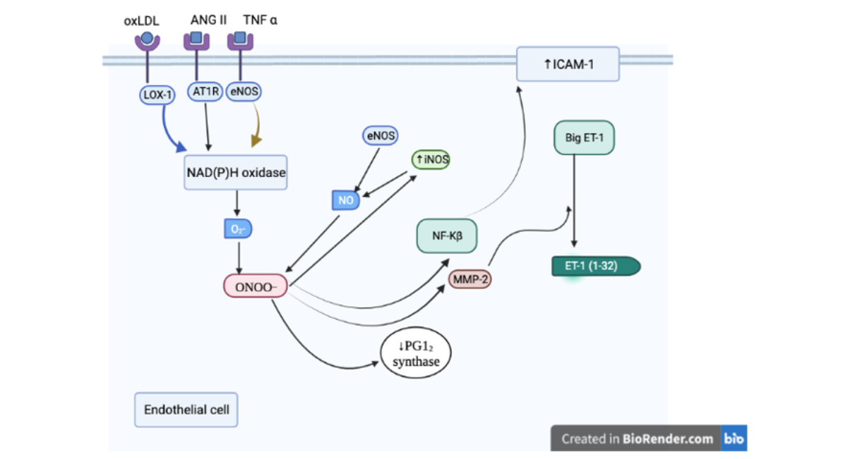
Figure 4: Oxidative stress mediated endothelial cell dysfunction in preeclampsia
Matrix Metalloproteinase
ROS and TNF-α have a role in the maternal vascular alterations associated with preeclampsia, which are released by activated neutrophils. Recent studies show that the potential mechanism for MMP-1 induction and activation may involve the generation of ROS and TNF- α by infiltrated neutrophils. This gives insight into how TNF-α and ROS modulate the expression of MMPs in vascular smooth muscle cells (VSMCs). Migration and local accumulation of leukocytes at the sites of inflammation are critical for the inflammatory response. To migrate, Leukocytes need to degrade the blood vessel basement membranes with proteases. The identification of MMP-1 as a mediator of neutrophil chemotaxis through liberation of IL-8 secretion and possibly collagen fragments by VSMCs provides insight into the mechanisms underlying vascular inflammation and, consequently, vascular dysfunction in women with preeclampsia. The signaling network triggered by neutrophils infiltrated into the vasculature during preeclampsia probably includes the expression of the thrombin receptor, PAR-1. VSMCs stimulated in vitro with activated neutrophils, TNF-α, or ROS elicit PAR-1 up-regulation. This G protein-coupled receptor plays a major role in orchestrating the interaction between coagulation and inflammation. The PAR-1 is a tethered ligand receptor that is activated by proteolytic cleavge of its extracellular domain. Activating proteases include thrombin, trypsin, factor Xa, factor XIIa/X and MMP-1. PAR-1 expression is increased in the vasculature of women with preeclampsia. Because PAR-1 is mostly confined to the endothelium in healthy human arteries, whereas during an inflammatory process, its expression is enhanced in regions associated with leukocyte influx. The Contact between infiltrating neutrophils and VSMCs could induce overexpression of PAR-1, which then may be activated by the MMP-1 present in the microenvironment, triggering a cascade of downstream events that result in vascular dysfunction in women with preeclampsia. The role of MMPs in vascular function includes regulation of vasoconstriction. Therefore, increasing levels of MMP-1 in preeclampsia seem to be central to the hypertensive disorder observed in these women. Therefore, vascular reactivity would be enhanced because of increased expression of both MMP-1 and PAR-1 in those with preeclampsia. Extracellular matrix remodeling has critical effects on vascular function and the consequent behavior of cells residing on or within it [24,44].
Generally, Activated neutrophils infiltrate the blood vessels. Once in the intimal space, neutrophils secrete TNF-α and ROS. This, in turn, stimulates VSMCs to secrete MMP-1 and IL-8. An increment of MMP-1 induces collagen breakdown, which possibly favors edema and proteinuria. The IL-8 and possibly collagen fragments induce recruitment of more neutrophils, favoring vascular inflammation. Active MMP-1 can play additional roles, including enhancement of neutrophil inflammatory response and vasoconstriction through the cleavage of PAR-1 receptor on endothelial cells or VSMCs [44].
The Role of the Renin-Angiotensin System in Preeclampsia
All major components of the renin-angiotensin system are present in the human placenta and related tissues. In this system, AT II can act in an autocrine/paracrine fashion. By mediating the inhibition of endothelial cell proliferation, it can stimulate angiogenesis and is capable of antiproliferative actions. The actions of AT II are mediated by at least two receptors, AT 1 and AT 2 receptor subtypes. The AT1 receptor mediates the vasoconstrictive and growth-promoting effects of AT II, the presence of which has been proven in the human placenta. On the other hand, AT 2 receptors, exerting an antiproliferative effect which opposes the growth action of AT 1 receptors, are practically not present. It implies it is unlikely that AT II mediates any significant action via the AT 2 receptors. Since renin, angiotensin converting enzyme, and AT receptor 1 are all expressed in and around remodeling spiral arteries. The known actions and presence of the renin-angiotensin system suggest that the local spiral artery renin-angiotensin system may play a role in the pregnancy-induced remodeling of these vessels [45].
Although the altered regulation of the renin-angiotensin system is implicated in the pathogenesis of preeclampsia, the reason behind these alterations is yet to be identified. However, AT II levels are reportedly decreased in preeclamptic women as compared with normotensive pregnant women. These patients show hypertension and renal dysfunction symptoms that could be attributed to excess AT1-receptor activation. Unfortunately, the exact cause of this excess activation remains elusive. However, the autoantibody secreted in preeclampsia that stimulates the AT1 receptor may explain these phenomena. It is possible that through excess AT1-receptor activation, the angiotensin II type I receptor agonistic autoantibody (AT1-AA) could produce preeclamptic phenotype symptoms in pregnant women. This implies AT1-AAs have a significant role in the PE syndrome [46].
AT1 receptors are activated on a variety of cell types; these autoantibodies could increase certain factors that lead to preeclamptic pathophysiology, such as endothelial cell dysfunction and vascular damage. The autoantibody may contribute to the pathogenesis of the disease in a variety of ways. It may induce the liberation of sFlt-1. Excess AT1-receptor activation may also lead to increased PAI-1. This excess PAI-1 could result in shallow trophoblast invasion and decreased extracellular matrix degradation and subendothelial and subepithelial fibrin deposits, thereby contributing to kidney damage. Increased glomerular fibrin deposition could result in a decrease in the kidney’s filtration ability. AT1-AAs increase PAI-1 in both the placenta and kidney and lead to decreased fibrinolysis and extracellular matrix breakdown, which ultimately participate in organ damage and symptoms associated with PE. The autoantibody also promotes the production of other factors, such as ROS [47].
CONCLUSION
Preeclampsia is a multifactorial disorder, and its etiology remains unclear. It is the maternal syndrome with a hallmark of vascular dysfunction, as a result of the release of trophoblastic factor following placental ischemia/hypoxia. Imbalance in the production of pro- and antiangiogenic factors, inflammation, altered renin angiotensin system, and oxidative stress within the endothelium are major contributors to endothelial dysfunction. Factors such as oxLDL, ANG II, and TNF-α may act both individually and synergistically to cause alteration of the maternal endothelial function. The release of these factors to the maternal vasculature leads to increased NO scavenging, which subsequently results in peroxynitrite formation, which leads to neutrophil infiltration. It further activates MMPs to induce vasoconstriction. The overall mechanisms suggest a role of trophoblastic factor in the pathogenesis of preeclampsia. The mechanisms by which the levels of such factors are altered, and their subsequent effect on the maternal endothelium, remain to be fully elucidated. Understanding of the mechanisms leading to vascular endothelial dysfunction and its feed-forward progression to preeclampsia is critically important for the prevention and treatment of this disorder.
FUTURE PERSPECTIVE
Oxidative stress, inflammation, altered renin angiotensin system, and angiogenic imbalance lead to maternal vascular dysfunction. It results from the interaction between the actions of placenta-derived factors and other circulating factors. Pharmacological manipulation of signaling pathway, which are central to inflammatory and vasoactive mechanisms, of lectin-like oxidized low-density lipoprotein receptor-1 (LOX-1), heme oxygenase-1 (HO-1) and Toll-like receptor (TLR) may provide novel strategies to target maternal vascular dysfunction selective pharmacological agents that target production and/or signaling of angiotensin receptor autoantibodies (AT1-AAs), Matrix metalloproteinases (MPPs), and cell-free nucleic acids (CpG DNA) will allow the investigation of their cellular mechanisms and vascular pathophysiology of preeclampsia. Finally, studies are needed to investigate the molecular mechanisms by which placenta-derived factors and other circulating factors facilitate derangements of maternal vascular biology during preeclampsia.
CREDIT AUTHORSHIP CONTRIBUTION STATEMENT
Authors contributed equally.
DATA AVAILABILITY
Since this is a review article, no new data were used.
FUNDING
This study did not receive funding from any governmental or non- governmental sources.
CONFLICT OF INTEREST
We have no conflict of interest.
ABBREVIATIONS
ACE Angiotensin-Converting Enzyme
AMH Anti-Mullerian Hormone
BMI Body Mass Index
CVD Cardiovascular Disease
DBP Diastolic Blood Pressure
DNA Deoxyribonucleic Acid
FFA Free Fatty Acid
FSH Follicle-Stimulating Hormone
GTRH Gonadotropins Releasing Hormone
HDL High-Density Lipoprotein
HFD High Fat Diet
HOMA-IR Homeostasis Model Assessment for Insulin Resistance
HPG Hypothalamic Pituitary Gonadotropic Axis
LDL Low-Density Lipoprotein
LH Luteinizing Hormone
MetS Metabolic Syndrome
REFERENCES
- Hauth JC, Ewell MG, Levine RJ, Esterlitz JR, Sibai B, et al. Pregnancy outcomes in healthy nulliparas who developed hypertension. Obstetrics & gynecology. 2000 Jan;95(1):24-8. [Google Scholar] [PubMed]
- Barton JR, Sibai BM. Prediction and prevention of recurrent preeclampsia. Obstetrics & Gynecology. 2008 Aug 1;112(2 Part 1):359-72. [Google Scholar] [PubMed]
- Roberts JM. Preeclampsia: what we know and what we do not know. InSeminars in perinatology 2000 Feb 1 (Vol. 24, No. 1, pp. 24-28). WB Saunders. [Google Scholar]
- Webster RP, Myatt L. Elucidation of the molecular mechanisms of preeclampsia using proteomic technologies. PROTEOMICS–Clinical Applications. 2007 Sep;1(9):1147-55. [Google Scholar] [PubMed]
- Vitoratos N, Hassiakos D, Iavazzo C. Molecular mechanisms of preeclampsia. Journal of Pregnancy. 2012;2012(1):298343. [Google Scholar] [PubMed]
- PALMER SK, Zamudio S, Coffin C, Parker S, Stamm E, et al. Quantitative estimation of human uterine artery blood flow and pelvic blood flow redistribution in pregnancy. Obstetrics & Gynecology. 1992 Dec 1;80(6):1000-6. [Google Scholar] [PubMed]
- Baumwell S, Karumanchi SA. Pre-eclampsia: clinical manifestations and molecular mechanisms. Nephron Clinical Practice. 2007 Jun 6;106(2):c72-81. [Google Scholar] [PubMed]
- Brennan LJ, Morton JS, Davidge ST. Vascular dysfunction in preeclampsia. 2014 Jan;21(1):4-14. [Google Scholar] [PubMed]
- Tal R. The role of hypoxia and hypoxia-inducible factor-1alpha in preeclampsia pathogenesis. Biology of reproduction. 2012 Dec 1;87(6):134-. [Google Scholar] [PubMed]
- Shamshirsaz AA, Paidas M, Krikun G. Preeclampsia, hypoxia, thrombosis, and inflammation. J Pregnancy. 2012;2012. [Google Scholar] [PubMed]
- Levine RJ, Maynard SE, Qian C, Lim KH, England LJ, et al. Circulating angiogenic factors and the risk of preeclampsia. New England journal of medicine. 2004 Feb 12;350(7):672-83. [Google Scholar] [PubMed]
- Nagai A, Sado T, Naruse K, Noguchi T, Haruta S, Yoshida S, et al. Angiogenic-induced hypertension: the molecular basis of signaling network. Gynecol Obstet Invest. 2012;73(2):89–98. [Google Scholar] [PubMed]
- He H, Venema VJ, Gu X, Venema RC, Marrero MB, et al. Vascular endothelial growth factor signals endothelial cell production of nitric oxide and prostacyclin through flk-1/KDR activation of c-Src. J Biol Chem. 1999;274(35):25130–5. [Google Scholar] [PubMed]
- Young BC, Levine RJ, Karumanchi SA. Pathogenesis of preeclampsia. Annu Rev Pathol Mech Dis. 2010;5:173–92. [Google Scholar] [PubMed]
- Nagai A, Sado T, Naruse K, Noguchi T, Haruta S, et al. Antiangiogenic-induced hypertension: the molecular basis of signaling network. Gynecol Obstet Invest. 2012;73(2):89–98. [Google Scholar] [PubMed]
- Hod T, Cerdeira AS, Karumanchi SA. Molecular mechanisms of preeclampsia. Cold Spring Harb Perspect Med. 2015;5(10):a023473. [Google Scholar] [PubMed]
- Kendall RL, Thomas KA. Inhibition of vascular endothelial cell growth factor activity by an endogenously encoded soluble receptor . Proc Natl Acad Sci. 1993;90(22):10705–9. [Google Scholar] [PubMed]
- Huckle WR, Roche RI. Post‐transcriptional control of expression of sFlt‐1, an endogenous inhibitor of vascular endothelial growth factor. J Cell Biochem. 2004;93(1):120–32. [Google Scholar] [PubMed]
- Sela S, Itin A, Natanson-Yaron S, Greenfield C, Goldman-Wohl D, et al. A novel human-specific soluble vascular endothelial growth factor receptor 1: cell type-specific splicing and implications to vascular endothelial growth factor homeostasis and preeclampsia. Circ Res. 2008;102(12):1566–74. [Google Scholar] [PubMed]
- Lapaire O, Holzgreve W, Oosterwijk JC, Brinkhaus R, Bianchi DW. Georg Schmorl on trophoblasts in the maternal circulation. Placenta. 2007;28(1):1–5. [Google Scholar] [PubMed]
- Baumwell S, Karumanchi SA. Pre-eclampsia: clinical manifestations and molecular mechanisms. Nephron Clin Pract. 2007;106(2):c72–81. [Google Scholar] [PubMed]
- Gandley RE, Tyurin VA, Huang W, Arroyo A, Daftary A, Harger G, et al. S-Nitrosoalbumin–Mediated Relaxation Is Enhanced by Ascorbate and Copper: Effects in Pregnancy and Preeclampsia Plasma. Hypertension. 2005;45(1):21–7. [Google Scholar] [PubMed]
- Sankaralingam S, Arenas IA, Lalu MM, Davidge ST. Preeclampsia: current understanding of the molecular basis of vascular dysfunction. Expert Rev Mol Med. 2006;8(3):1–20. [Google Scholar] [PubMed]
- Ignarro LJ. Nitric oxide. A novel signal transduction mechanism for transcellular communication. 1990;16(5):477–83. [Google Scholar] [PubMed]
- Xia Y, Dawson VL, Dawson TM, Snyder SH, Zweier JL. Nitric oxide synthase generates superoxide and nitric oxide in arginine-depleted cells, leading to peroxynitrite-mediated cellular injury. Proc Natl Acad Sci. 1996;93(13):6770–4. [Google Scholar] [PubMed]
- Baker PN, Davidge ST, Roberts JM. Plasma from women with preeclampsia increases endothelial cell nitric oxide production. Hypertension. 1995;26(2):244–8. [Google Scholar] [PubMed]
- Roggensack AM, Zhang Y, Davidge ST. Evidence for peroxynitrite formation in the vasculature of women with preeclampsia. 1999;33(1):83–9. [Google Scholar] [PubMed]
- Busse R, Edwards G, Félétou M, Fleming I, Vanhoutte PM, et al. EDHF: bringing the concepts together. Trends Pharmacol Sci. 2002;23(8):374–80. [Google Scholar] [PubMed]
- Pascoal IF, Lindheimer MD, Nalbantian-Brandt C, Umans JG. Preeclampsia selectively impairs endothelium-dependent relaxation and leads to oscillatory activity in small omental arteries. J Clin Invest. 1998;101(2):464–70. [Google Scholar] [PubMed]
- Luksha L, Kublickiene K. Implications for endothelium-derived hyperpolarizing factor (EDHF) in women’s cardiovascular health. Curr Womens Health Rev. 2005;1(1):67–78. [Google Scholar]
- Saito S, Shiozaki A, Nakashima A, Sakai M, Sasaki Y. The role of the immune system in preeclampsia. Mol Aspects Med. 2007;28(2):192–209. [Google Scholar] [PubMed]
- Takacs P, Kauma SW, Sholley MM, Walsh SW, Dinsmoor MJ, Green K. Increased circulating lipid peroxides in severe preeclampsia activate NF-κB and upregulate ICAM-1 in vascular endothelial cells. FASEB J. 2001;15(2):279–81. [Google Scholar] [PubMed]
- Zhang Q, Raoof M, Chen Y, Sumi Y, Sursal T, Junger W, et al. Circulating mitochondrial DAMPs cause inflammatory responses to injury. Nature. 2010;464(7285):104. [Google Scholar] [PubMed]
- Hahn S, Giaglis S, Buser A, Hoesli I, Lapaire O, et al. Cell-free nucleic acids in (maternal) blood: any relevance to (reproductive) immunologists? J Reprod Immunol. 2014;104:26–31. [Google Scholar] [PubMed]
- Scharfe-Nugent A, Corr SC, Carpenter SB, Keogh L, Doyle B, et al. TLR9 provokes inflammation in response to fetal DNA: mechanism for fetal loss in preterm birth and preeclampsia. J Immunol. 2012;188(11):5706–12. [Google Scholar] [PubMed]
- Zhong XY, Holzgreve W, Hahn S. The levels of circulatory fetal DNA in maternal plasma are elevated prior to the onset of preeclampsia. Hypertens Pregnancy. 2002;21(1):77–83. [Google Scholar] [PubMed]
- Rani N, Dhingra R, Arya DS, Kalaivani M, Bhatla N, Kumar R. Role of oxidative stress markers and antioxidants in the placenta of preeclamptic patients. J Obstet Gynaecol Res. 2010;36(6):1189–94. [Google Scholar] [PubMed]
- Roggensack AM, Zhang Y, Davidge ST. Evidence for peroxynitrite formation in the vasculature of women with preeclampsia. 1999;33(1):83–9. [Google Scholar] [PubMed]
- Brennan LJ, Morton JS, Davidge ST. Vascular dysfunction in preeclampsia. Microcirculation. 2014;21(1):4–14. [Google Scholar] [PubMed]
- Sankaralingam S, Xu Y, Sawamura T, Davidge ST. Increased lectin-like oxidized low-density lipoprotein receptor-1 expression in the maternal vasculature of women with preeclampsia: role for peroxynitrite. 2009;53(2):270–7. [Google Scholar] [PubMed]
- Sun Y, Chen X. Ox‐LDL‐induced LOX‐1 expression in vascular smooth muscle cells: role of reactive oxygen species. Fundam Clin Pharmacol. 2011;25(5):572–9. [Google Scholar] [PubMed]
- Cudmore M, Ahmad S, Al-Ani B, Fujisawa T, Coxall H, et al. Negative regulation of soluble Flt-1 and soluble endoglin release by heme oxygenase-1. Circulation. 2007;115(13):1789–97. [Google Scholar] [PubMed]
- Estrada-Gutierrez G, Cappello RE, Mishra N, Romero R, Strauss III JF, Walsh SW. Increased expression of matrix metalloproteinase-1 in systemic vessels of preeclamptic women: a critical mediator of vascular dysfunction. Am J Pathol. 2011;178(1):451–60. [Google Scholar] [PubMed]
- Tálosi G, Endreffy E, Túri S, Németh I. Molecular and genetic aspects of preeclampsia: state of the art. Mol Genet Metab. 2000;71(4):565–72. [Google Scholar] [PubMed]
- Wallukat G, Homuth V, Fischer T, Lindschau C, Horstkamp B, et al. Patients with preeclampsia develop agonistic autoantibodies against the angiotensin AT1 receptor. J Clin Invest. 1999;103(7):945–52. [Google Scholar] [PubMed]
- Irani RA, Xia Y. Renin angiotensin signaling in normal pregnancy and preeclampsia. In: Seminars in nephrology. Elsevier; 2011. p. 47–58. [Google Scholar] [PubMed]