




 Download PDF
ABSTRACT
Cervical cancer presents a significant global health challenge, underscoring the importance of early detection to prevent and manage the disease. This study utilizes a dataset comprising 1500 JPG images (224x224x3) of cervical precancerous lesions, sourced from the IARC cervical cancer image bank. The images are classified as positive, negative, or suspicious for cancer. Employing Python, TensorFlow, Keras, OpenCV, NumPy, and Pandas, the research applies various preprocessing techniques, including data augmentation and CLAHE. The model architecture incorporates a CNN with Squeeze-and-Excitation blocks for feature refinement. The dataset is split into an 80:20 ratio, and the model undergoes 100 epochs of training with a batch size of 32 and a learning rate of 0.001. Evaluation metrics encompass accuracy, precision, recall, F-Measure, confusion matrices, and ROC curves. Preprocessing significantly boosts classification accuracy compared to a model without preprocessing. Using k-fold cross-validation (k=3) on test data before preprocessing, the model achieves an accuracy of 94.24%. While initial findings reveal promising ROC-AUC scores, erratic accuracy and loss graphs underscore the necessity for preprocessing. Misclassifications in validation and test sets are attributed to data limitations. While after preprocessing techniques, such as CLACHE, dataset expansion, and increased epochs, the accuracy is elevated to 100%, with ROC-AUC scores reaching 100% for all classes. The confusion matrix indicates no misclassifications post-preprocessing, while the classification report showcases optimal precision, recall, F1-score, and accuracy across datasets, highlighting the efficacy of preprocessing techniques. This study underscores the pivotal role of preprocessing techniques in enhancing the accuracy of detecting cervical precancerous cells. It emphasizes their significance in early lesion identification and cervical cancer risk screening.
Keywords: Cervical Cancer: Squeeze-and-Excitation Blocks: Preprocessing Techniques
INTRODUCTION
Cancer, along with cardiovascular disease, diabetes, and chronic respiratory diseases, is a major noncommunicable disease contributing to over 60% of global mortality annually. It is estimated that cancer causes over 7.9 million deaths worldwide each year, accounting for nearly 13% of all deaths[1-6]. Among women in developing countries, breast and cervical cancers are the most prevalent, with approximately 882,900 and 444,500 new cases annually. These cancers lead to the deaths of more than 324,300 and 230,400 women each year respectively [6]. In less developed countries, it ranks as the second most diagnosed cancer and the third leading cause of cancer death among female [1].
Cervical cancer begins with abnormal cell growth in the cervix; the lower part of the uterus connected to the vagina. It's a major cause of death for women, with the highest occurrence in 23 countries and the highest death rates in 36 countries, primarily in sub-Saharan Africa, Melanesia, South America, and South-Eastern Asia [2].
According to the 2015 Ethiopian cervical cancer prevention and control guidelines, cervical cancer is the most prevalent reproductive organ cancer and a leading cause of death in low and middle-income countries (LMICs), such as Ethiopia [6]. The updated 2021 guidelines state that cervical cancer ranks as the fourth most common cancer globally for women, with around 569,847 new cases and over 311,000 deaths annually(2021).Cervical cancer is preventable, unlike other reproductive organ cancers. Screening programs can significantly reduce morbidity and mortality associated with this cancer. In high-income countries, regular Pap smear screenings lower the risk of invasive cervical cancer by detecting precancerous changes. Screening targets individuals at risk, aiming to identify those with a high probability of developing the disease(2021).
Visual inspection with acetic acid (VIA) and Lugol’s iodine (VILI) are two visual methods for cervical cancer screening. The new screening method adopts HPV DNA testing to enhance screening. Colposcopy is crucial for visually inspecting cervical epithelial tissue to prevent cervical cancer. However, artificial vision screening poses challenges such as missed diagnoses, low efficiency, and diversity [3].
Medical image processing and AI contribute to the analysis of malignant cells, becoming cost-effective and less time-consuming with new techniques. They are increasingly preferred over traditional methods like Pap smear, Colposcopy, and Cervicography due to their objectivity. While they cannot fully replace expert doctor evaluations, they greatly assist them [4].
Deep learning models draw inspiration from biological nervous systems but differ significantly from the human brain in function and structure. They are applied in computer vision, bioinformatics, and other fields, often achieving results comparable to or surpassing human experts [5]. Research in the field of deep learning applied to diagnosing cervical cancer yielded promising results. While yielding promising results, still some research gaps including. refining methods for acquiring colposcopy raw images, collecting multicenter data and conducting comprehensive research for clinical adaptation, and enhancing model effectiveness by incorporating additional metrics [6]. Further optimization of data augmentation methods, in-depth studies on cell integrity for improved classification accuracy, addressing challenges related to dataset diversity, scanner types, patient ages, and background noise, and exploring additional research avenues to enhance model robustness and reliability are also essential [7]. Studies could assess the screening accuracy of the developed algorithm in clinical settings and explore combining it with other screening methods for improved early detection and screening outcomes for cervical cancer [8]. These findings stimulate further research on computer-aided diagnosis for more efficient and accurate screening approaches. The Squeeze-and-Excitation (SE) block enhances neural networks by modeling channel-wise dependencies, recalibrating feature responses to emphasize informative features and suppress less useful ones. Introduced by Jie Hu et al., it boosts performance in computer vision tasks by enabling networks to focus on important features. Effective across CNN architectures, the SE block improves accuracy and generalization by adaptively emphasizing informative features. Its lightweight design facilitates efficient integration into existing architectures, enhancing performance with low latency [9]. Here, we propose a research study to develop a model for classifying cervical precancerous lesions using a squeezed convolutional neural network. The study aims to achieve several specific objectives, applying preprocessing techniques to cervical precancerous lesion images, designing the proposed architecture for these images, building a SECNN model for lesion classification, and evaluating the model's performance using performance metrics.
Literature Review
Cervical cancer is a leading cause of cancer among women, particularly in developing countries where screening services and specialist care are limited. Common screening methods include Visual inspection is used to screen for cervical cancer after the application of acetic acid (VIA), histopathology, Papanicolaou (Pap), and human papillomavirus (HPV) examinations. To enhance early detection, researchers are developing vision transformer–based deep learning models for accurate classification of cervical precancerous lesions. Additionally, they seek to develop a high-precision cervical cell classification model using the DCCL dataset to enable reliable diagnosis and analysis of cervical cancer [10]. Another study focuses on developing a deep learning-based algorithm for the detection and classification of precancerous lesions and cervical cancer, with primary objectives to establish an accurate screening method and evaluate model performance using multiple metrics [11]. The study utilized a vision transformer with shifted patch tokenization to distinguish between three cervical pre-cancerous types. Training and testing on 8215 colposcopy images from the mobile-ODT dataset showed a 91% accuracy, outperforming previous models [12]. Additionally, they extracted 8619 distinctive cervical cytological images, classified them into four categories, and proposed a self-supervised data augmentation technique. Their approach surpassed traditional convolutional neural network models and achieved significant improvements in accuracy compared to mainstream models, reaching a 10.43% improvement over a Swin transformer and an 8.85% improvement over Beit [13]. The study employed a cervical cancer screening dataset with 1,189 patients and 6,996 images, implementing data augmentation techniques for model robustness. Convolutional Neural Network algorithms in PyTorch were utilized with specific parameters and trained for 100 cycles. The model achieved an accuracy of approximately 83% in detecting and classifying precancerous lesions and cervical cancer, with reported precision, recall, and F1-score [14]. Research gaps include refining methods for acquiring colposcopy raw images, collecting multicenter data and conducting comprehensive research for clinical adaptation, and enhancing model effectiveness by incorporating additional metrics [15]. Further optimization of data augmentation methods, in-depth studies on cell integrity for improved classification accuracy, addressing challenges related to dataset diversity, scanner types, patient ages, and background noise, and exploring additional research avenues to enhance model robustness and reliability are also essential [16]. Studies could assess the screening accuracy of the developed algorithm in clinical settings and explore combining it with other screening methods for improved early detection and screening outcomes for cervical cancer. Previously researchers have developed ML-based colposcopy models for classifying cervical neoplasms using the CIN and LAST systems, evaluating their efficacy in identifying lesions requiring biopsies.Additionally, CerviCARE AI, an AI analysis software, aims to distinguish low-grade and high-grade lesions using Tele-cervicography images. Another group focuses on developing an efficient algorithm for segmenting cervical lesions in colposcopic images to enhance sensitivity for early detection and timely medical intervention in cervical cancer cases. Researchers utilized retrospective colposcopic photographs of cervical lesions verified by pathology. Inclusion criteria: Women ≥ 18 years, non-pregnant, no cervical surgery history, with Pap test results. Lesion classification used CIN model and LAST system. Training and test datasets constructed (85%:15%). Dataset pre-processing involved augmentation and normalization. CNN models trained (Inception-Resnet-v2 and Resnet-152) with stages including pre-trained model loading, layer unfreezing, cyclic training, and differential learning rates. CAM implemented for region of interest detection. Inception-Resnet-v2 achieved 48.6 ± 1.3% accuracy for CIN, and Resnet-152 achieved 51.7 ± 5.2%. For LAST, accuracies were 71.8 ± 1.8% and 74.7 ± 1.8%, respectively. Resnet-152 showed AUC of 0.781 ± 0.020 for CIN and 0.708 ± 0.024 for LAST. Efficient detection of biopsy-requiring lesions (AUC 0.947 ± 0.030). Results suggest AI's potential for automated colposcopic photograph reading.Multicenter retrospective analysis assessed CerviCARE AI performance in Tele-cervicography image analysis. Designed to distinguish low-grade and high-grade lesions from HR-HPV infection. Compared with Pap test, LBC, VIA, and HPV DNA testing, CerviCARE AI achieved 98% sensitivity for high-risk groups and 95.5% specificity. AI system can accurately identify cervical precancerous lesions, enhancing cervical cancer screening.Cervical lesion segmentation method comprised three stages: Faster R-CNN for cervix region location, CLS-Net for lesion segmentation, and mapping results to original image size. Evaluated on 10,300 cervigram images, outperforming existing techniques with higher sensitivity (HR 0.9304) and Dice Similarity Coefficient (DSC 0.7802). High accuracy may lead to earlier cervical cancer detection and improved treatment outcomes.Address data imbalance, enhance specificity, reduce false positives, standardize image characteristics, and integrate clinical parameters to improve machine learning models for colposcopy.However, research is needed to validate CerviCARE AI's clinical utility, integrate it into existing protocols, evaluate real-world performance, explore cost-effectiveness, and scalability.Additional work should involve testing on larger datasets, exploring limitations due to lighting and image quality, extending features to improve lesion detection accuracy, and assessing feasibility in clinical settings.
MATERIALS AND METHODS
Dataset
In this study the cervical precancerous lesion images from the IARC cervical cancer image bank's online data repository, which were in .JPG format. The images, initially of various sizes, were resized to 224x224x3 pixels and contains three classes: negative (187), positive (190), and suspicious for cancer (40). Later, the numbers increased to negative (488), positive (489), and suspicious for cancer (491) using data augmentation techniques.
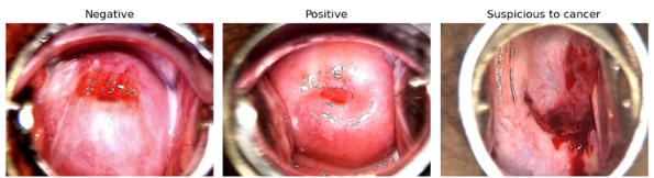
Figure 1 : A sample of three different types of cervical cancerous conditions from the dataset
Tools and materials
Python, distinguished from MATLAB and C++ for its popularity in scientific computing and machine learning, excels in readability, versatility, and library support. Its extensive ecosystem includes NumPy, SciPy, Pandas, and Scikit-learn, bolstering its dominance in machine learning, optimization, and GPU-accelerated computing. In this study we utilized Python, along with TensorFlow and Keras, for image processing tasks, supported by OpenCV, NumPy, and Pandas. The experimental setup comprised an HP laptop running Windows 11 with an Intel(R) Core (TM) i5-8250U CPU, 8 GB RAM, and a 1TB hard drive. Documentation was done using Microsoft Word 2019, aided by the Zotero referencing tool.
Data Preprocessing
Previous studies commonly utilized several preprocessing techniques. Normalization, achieved through min-max normalization, aimed to reduce image differences.Data augmentation, including customized approaches such as six-fold rotation for less frequent classes and various flips, addressed data imbalance issues and enhanced the dataset. Image sharpening was employed to improve edge contrast and reduce noise, enhancing overall image clarity.Contrast Limited Adaptive Histogram Equalization (CLAHE) was used to improve contrast across the image while limiting noise, resulting in a clearer and more uniform image.Various data splitting techniques are vital in Computer Vision to enhance model robustness and fairness. Random sampling shuffles datasets for training, validation, or testing, though it can introduce bias in imbalanced datasets. Stratified sampling preserves class distribution, mitigating bias. Cross-validation, particularly Stratified K-Fold, repeatedly trains and evaluates models on diverse samples for reliable performance assessment. Optimal split percentages, like 80:20,70:30, balance specific requirements and data constraints, necessitating adjustment based on individual circumstances(Train Test Validation Split, n.d.).
MODEL TRAINING
A Convolutional Neural Network (CNN) model is specifically developed for the classification of cervical cancer images, employing sophisticated techniques and methodologies to enhance its classification accuracy. The Squeeze-and-Excitation (SE) block is integrated to boost the neural network's representational capacity by explicitly modeling interdependencies among channels within the intermediate feature maps derived from convolutional layers. The construction of the CNN model using TensorFlow's Keras API is systematic. It initiates with defining an input layer with dimensions of 224x224x3 to match the input image size. Sequentially, a series of convolutional layers are applied, each configured with distinct filter sizes, kernel sizes, activation functions, and padding schemes. Max-pooling operations are applied to down-sample the feature maps. Notably, Squeeze-and-Excitation blocks are inserted after specific convolutional layers to further refine feature representation. To prevent overfitting, a dropout layer with a rate of 0.5 is introduced, followed by a flattening layer to prepare the output for subsequent fully connected layers. Dense layers with 512 units and 'relu' activation functions are employed for additional feature extraction and transformation. The output layer, with 512 units corresponding to the three classes, utilizes SoftMax activation for multi-class classification. During the model compilation stage, key hyperparameters are configured, including the Adam optimizer with a learning rate of 0.001, categorical cross entropy loss function, and accuracy metric. The training regimen comprises 100 epochs, a batch size of 32, and an 80%:20% training-testing split ratio. The described CNN model architecture is meticulously designed and equipped with advanced components to effectively classify cervical cancer images.

Figure 2 : Proposed squeezed CNN model architecture
MODEL EVALUATION
The model's classification performance was assessed using various metrics including accuracy, precision, recall, F-Measure, confusion matrices, and ROC curve values. F-Measure, sensitive to overlaps between predicted segmentation and ground truth, is widely utilized in computer vision and Medical Imaging Segmentation (MIS), offering a balanced view of sensitivity and precision. Sensitivity focuses on true positive detection, crucial in medical evaluation, while specificity gauges the model's ability to detect background classes accurately. Accuracy, also known as Rand index, can be misleading in MIS due to class imbalance. The ROC curve illustrates a classifier's diagnostic ability across different discrimination thresholds, with the AUC indicating overall performance higher AUC values signify superior classifier performance, commonly used in medical contexts for diagnostic tests and clinical trials.The following equation describes the evaluation metrics:
Accuracy = (TP + TN) / (TP + TN + FN + FP)
AUC = 1 - 0.5 * (FP / (FP + TN) + FN / (FN + TP))
F-measure = 2 * (Precision * Sensitivity) / (Precision + Sensitivity)
Sensitivity = TP / (TP + FN)
Specificity = TN / (TN + FP)
EXPERIMENTAL RESULT
Experimental result before preprocessing
We conducted an experiment using images resized to 224x224x3 pixels, employing 50 epochs with a learning rate of 0.001 and a batch size of 32. The dataset was split into 80% training and 20% testing. Below are some of the resulting metrics.
Performance evaluation on test data from Experimental results before to preprocessing using k-fold cross-validation (k=3)
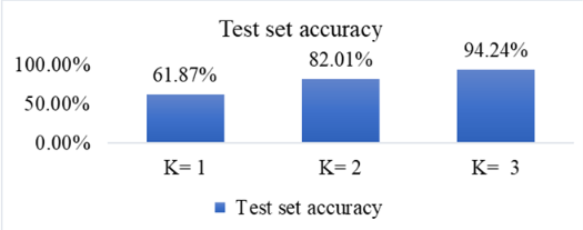
Figure 3 : Model accuracy of test set before preprocessing using k-fold cross-validation with k=3
Graphs depicting the ROC-AUC curve score and the model's loss and accuracy before preprocessing in the experimental results
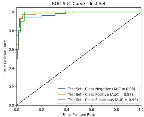
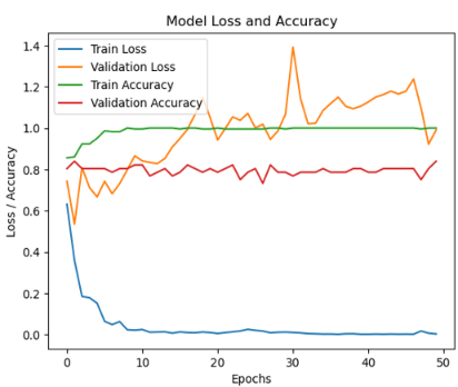
Figure 4 : Graphs depicting the ROC-AUC curve score and the model's loss and accuracy before preprocessing in the experimental results
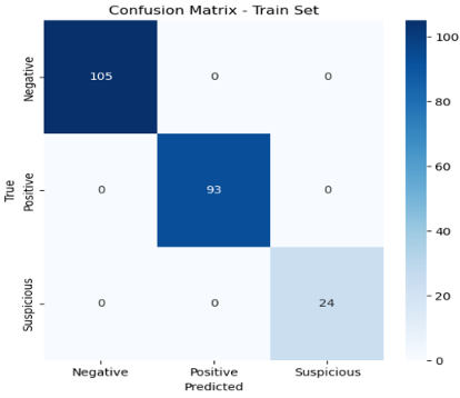
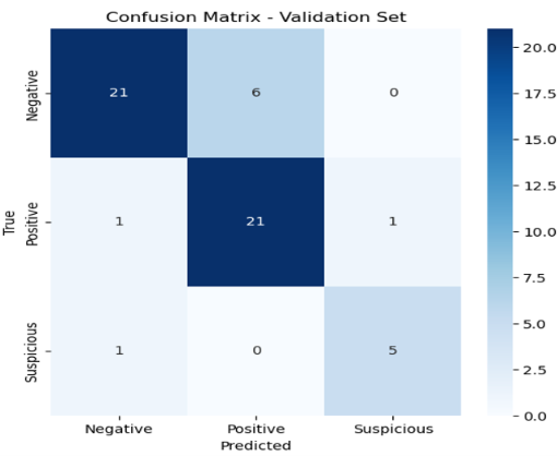
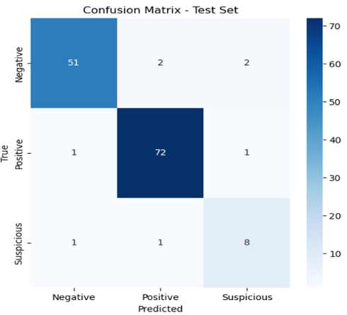
Figure 5 : confusion matrix before preprocessing in the experimental results for training, validation & test set
Report on classification results for the test set before preprocessing
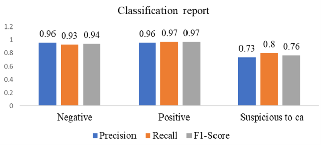
Figure 6 : Report on classification results for the test set before preprocessing
Experiment after preprocessing
Based on the initial results observed prior to preprocessing, it became apparent that the study required some preprocessing steps. Consequently, we implemented CLAHE, augmented the dataset, and extended the epoch to 100, resulting in a significant improvement in performance.
Accuracy of the experimental findings following preprocessing for the training, validation, and test sets
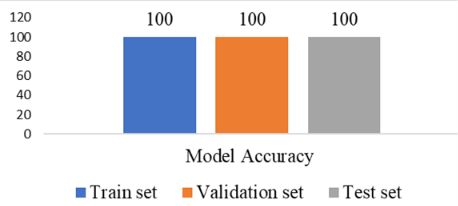
Figure 7 : model accuracy of the experimental findings following preprocessing for the training, validation, and test sets
Graphs depicting the ROC-AUC curve score and the model's loss and accuracy before preprocessing in the experimental results
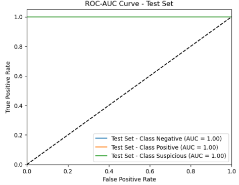
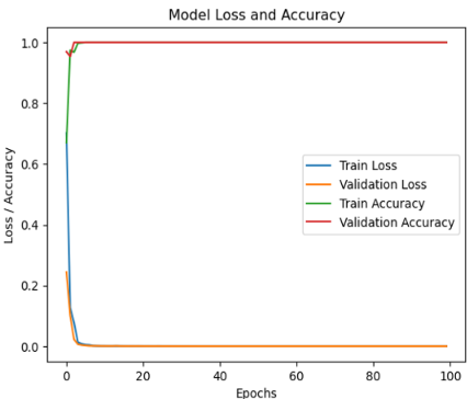
Figure 8 : Graphs depicting the ROC-AUC curve score and the model's loss and accuracy before preprocessing
Confusion matrix before preprocessing in the experimental results
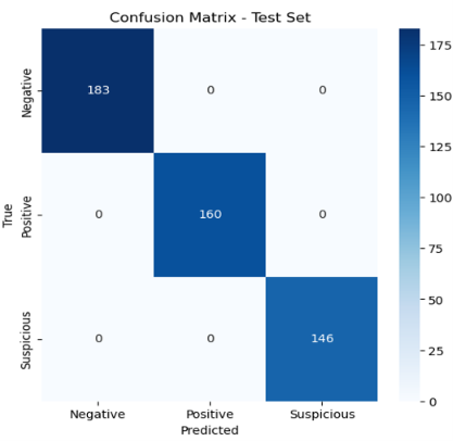
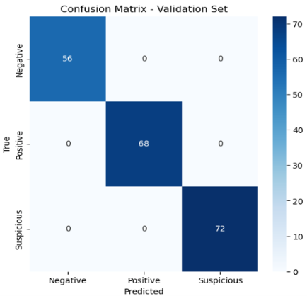
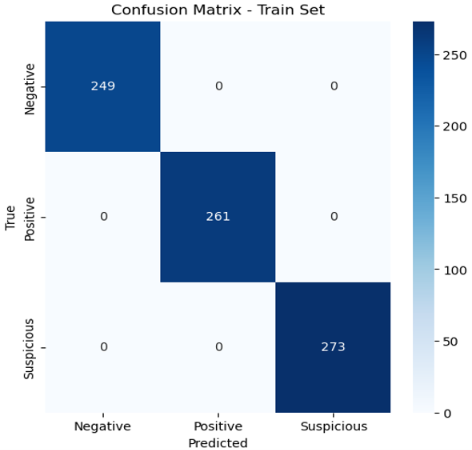
Figure 9 : Confusion matrix after preprocessing in the experimental results of train, validation & test set
Classification Report
|
Set |
Class |
Precision |
Recall |
F1-Score |
Accuracy |
|
Train Set |
Negative |
1.00 |
1.00 |
1.00 |
1.00 |
|
Positive |
1.00 |
1.00 |
1.00 |
||
|
Suspicious |
1.00 |
1.00 |
1.00 |
||
|
Validation Set |
Negative |
1.00 |
1.00 |
1.00 |
1.00 |
|
Positive |
1.00 |
1.00 |
1.00 |
||
|
Suspicious |
1.00 |
1.00 |
1.00 |
||
|
Test Set |
Negative |
1.00 |
1.00 |
1.00 |
1.00 |
|
Positive |
1.00 |
1.00 |
1.00 |
||
|
Suspicious |
1.00 |
1.00 |
1.00 |
Table 1 : Classification Report after preprocessing
RESULTS AND DISCUSSION
The performance evaluation on test data, using experimental outcomes prior to preprocessing, employs k-fold cross-validation (k=3). Results show that with k-1 folds, testing accuracy is 61.87%; with k-2 folds, it increases to 82.01%; and with k-3 folds, it reaches 94.24%, as shown in Figure 3. Figure 4 depicts the ROC-AUC curve score, model loss, and accuracy prior to preprocessing in the experimental findings. The AUC score for negative and positive classes is 98, with an AUC score of 99 for suspicious to cancer. Nonetheless, the graphs illustrating the model's loss and accuracy show some noise, suggesting a necessity to mitigate disturbances in the data, despite the promising results observed with the training data. Figure 5 illustrates the confusion matrix from the initial experimental results, depicting the training, validation, and test sets before preprocessing. The training set shows no misclassifications, suggesting satisfactory performance. However, the validation and test sets reveal some misclassifications, likely due to the dataset's limited size and the absence of preprocessing procedures. The test set results, shown in Figure 6 before preprocessing, reveal performance metrics including Precision, Recall, and F1-Score across different classes. For the "Negative" class, Precision, Recall, and F1-Score are 0.96, 0.93, and 0.94, respectively. Similarly, the "Positive" class displays a Precision of 0.96, Recall of 0.97, and an F1-Score of 0.97. Notably, the "Suspicious to cancer (ca)" class performs with a Precision of 0.73, Recall of 0.80, and an F1-Score of 0.76. However, these results prompt further examination and analysis to understand classification performance and identify areas for improvement. Upon reviewing the preliminary findings pre-processing, it was evident that the study necessitated certain preprocessing measures. Previous researchers trained and tested a model on 8215 colposcopy images from the mobile-ODT dataset. Testing on 30% of the dataset yielded a 91% accuracy, indicating strong generalization capability. The study used the DCCL dataset with 1167 cervical cytology specimens (ages 32 to 67) to train and validate an algorithm. The final classification accuracy on the DCCL dataset was 8.85%. Prior CNN models achieved 81.23% and 81.38% accuracy on test data, with an AUC score of 0.99, showing strong performance in AI-based colposcopy image classification, suitable for clinical use. Accordingly, we introduced CLACHE, expanded the dataset, and prolonged the epoch to 100, yielding a notable enhancement in performance, culminating in 100% accuracy across the training, validation, and test sets, as illustrated in Figure 7. The ROC-AUC scores, alongside the post-preprocessing model's loss and accuracy metrics, present significant insights. Notably, the AUC scores for each class achieve a flawless 100%, highlighting outstanding performance. The ROC curves visually depict the high-quality graph representations, affirming the model's robust discriminatory capabilities across diverse classes. Additionally, the model's accuracy and loss graph indicate an upward trend in accuracy and a simultaneous decrease in loss, attributed to the application of preprocessing techniques, as illustrated in Figure 8. Following the preprocessing tasks' completion, an in-depth examination of the confusion matrix within the experimental results indicates the absence of any misclassifications in both the train and validation sets, as well as the test set. This commendable outcome can be attributed to the thorough preprocessing procedures, as depicted in Figure 9. The classification report in Table 3 exhibits excellent performance across all datasets after preprocessing. Precision, recall, F1-score, and accuracy metrics for each class, Negative, Positive, and Suspicious are consistently optimal in the training, validation, and test sets, indicating a flawless classification scenario. These results highlight the effectiveness of the preprocessing techniques utilized, leading to reliable model predictions across diverse datasets.
CONCLUSION
The research findings suggest that employing the Squeezed Convolutional Neural Network model alongside specific preprocessing techniques yields increased accuracy in classifying cervical pre-cancerous cells. This underscores the potential effectiveness of preprocessing methods in early lesion detection. The outcomes underscore the significance of integrating preprocessing methodologies in cervical pre-cancerous cell screening and detection, facilitating the timely identification of patients vulnerable to cervical cancer.
RECOMMENDATION
It is advisable for medical practitioners to incorporate the proposed compressed CNN model along with preprocessing methods into their clinical decision-making protocols and cervical pre-cancerous lesion screenings. Recent medical investigations applying preprocessing methods to images have yielded progress in various medical domains, and the findings of this study offer significant perspectives on the efficacy of such techniques in enhancing cervical cancer prevention endeavors. Furthermore, future studies could investigate alternative preprocessing methods, expand the range of positive cancer class types and grades, and diversify datasets to enhance the diagnostic precision of the proposed model even further. Deployment and utilization of the model in clinical practice can facilitate real-world applications.
REFERENCES
- Cho BJ, Choi YJ, Lee MJ, Kim JH, Son GH, Park SH, Kim HB, Joo YJ, Cho HY, Kyung MS, Park YH. Classification of cervical neoplasms on colposcopic photography using deep learning. Scientific reports. 2020 Aug 12;10(1):13652. [Crossref] [Google Scholar] [PubMed]
- Darwish M, Altabel MZ, Abiyev RH. Enhancing cervical pre-cancerous classification using advanced vision transformer. Diagnostics. 2023 Sep 8;13(18):2884. [Crossref] [Google Scholar] [PubMed]
- Dash S, Acharya BR, Mittal M, Abraham A, Kelemen A, editors. Deep learning techniques for biomedical and health informatics. Cham, Switzerland: Springer International Publishing; 2020. [Google Scholar]
- Fang S, Yang J, Wang M, Liu C, Liu S. An improved image classification method for cervical precancerous lesions based on ShuffleNet. Computational intelligence and neuroscience. 2022;2022(1):9675628. [Crossref] [Google Scholar][PubMed]
- Ghoneim A, Muhammad G, Hossain MS. Cervical cancer classification using convolutional neural networks and extreme learning machines. Future Generation Computer Systems. 2020 Jan 1;102:643-9. [Google Scholar]
- Ethiopia F. Guideline for Cervic Cancer Prevention and Control in Ethiopia. Addis Ababa: Federal Ministry of Health Ethiopia. 2015. [Google Scholar]
- Hu SY, Zhao XL, Zhang Y, Qiao YL, Zhao FH. [Interpretation of" WHO guideline for screening and treatment of cervical pre-cancer lesions for cervical cancer prevention. Zhonghua yi xue za zhi. 2021 Sep 1;101(34):2653-7. [Crossref] [Google Scholar] [PubMed]
- Luo YM, Zhang T, Li P, Liu PZ, Sun P, Dong B, Ruan G. MDFI: multi-CNN decision feature integration for diagnosis of cervical precancerous lesions. IEEE Access. 2020 Feb 10;8:29616-26. [Google Scholar]
- Manaswi NK, Manaswi NK, John S. Deep learning with applications using python. Berkeley, CA, USA: Apress; 2018. [Google Scholar]
- Müller D, Soto-Rey I, Kramer F. Towards a guideline for evaluation metrics in medical image segmentation. BMC research notes. 2022 Jun 20;15(1):210. [Crossref] [Google Scholar] [PubMed]
- Ouh YT, Kim TJ, Ju W, Kim SW, Jeon S, Kim SN, Kim KG, Lee JK. Development and validation of artificial intelligence-based analysis software to support screening system of cervical intraepithelial neoplasia. Scientific reports. 2024 Jan 23;14(1):1957. [Crossref] [Google Scholar] [PubMed]
- Raschka S, Patterson J, Nolet C. Machine learning in python: Main developments and technology trends in data science, machine learning, and artificial intelligence. 2020 Apr 4;11(4):193. [Google Scholar]
- Tang J, Zhang T, Gong Z, Huang X. High precision cervical precancerous lesion classification method based on ConvNeXt. Bioengineering. 2023 Dec 15;10(12):1424. [Crossref] [Google Scholar] [PubMed]
- Baheti P. Train test validation split: How to & best practices [2023]. URl: https://www. v7labs. com/blog/train-validation-test-set. 2021. [Google Scholar]
- Vasu PK, Gabriel J, Zhu J, Tuzel O, Ranjan A. Mobileone: An improved one millisecond mobile backbone. In2023 IEEE/CVF Conference on Computer Vision and Pattern Recognition (CVPR) 2023 Jun 17 (pp. 7907-7917). IEEE. [Google Scholar]
- Yu H, Fan Y, Ma H, Zhang H, Cao C, Yu X, Sun J, Cao Y, Liu Y. Segmentation of the cervical lesion region in colposcopic images based on deep learning. Frontiers in oncology. 2022 Aug 3;12:952847. [Crossref] [Google Scholar] [PubMed]